Pelican-Cancer-Foundation-logos
Donate Now
Search
Pelican-Cancer-Foundation-logos

Dedicated to helping patients with bowel and liver cancer live well for longer

Donate Now
Search
  • Home
  • About us
    • About us
    • What we do
      • Our achievements
      • The Future
    • Who we are
      • Trustees
      • Our Team
      • Patrons
      • Annual Accounts
    • Partnerships
    • Vacancies
    • News
  • Education
    • Pelican OReCO
      • OReCO On-line Resources
      • OReCO Partners & Sponsors
      • Pelican OReCO Workshops
    • Pelican IMPACT
      • IMPACT – Completion & Evaluation
      • Workshops
      • IMPACT – Online resources
      • IMPACT – Partners and Sponsors
    • SPECC
      • SPECC – Completion & Evaluation
      • Workshops
      • SPECC – Online resources
      • SPECC – Partners and Sponsors
    • LOREC
    • TIPTOP
      • Workshops
      • TIPTOP – Online resources
      • Bob’s story (prostate cancer)
      • Andrew’s story (prostate cancer)
      • Alan’s story (prostate cancer)
      • Raymond’s story (prostate cancer)
    • LARS
    • Sponsors
      • Become a sponsor
    • Request a course or topic
    • Give us feedback
    • Previous courses
  • Films
  • Research
    • MERCURY 3
    • Research strategy
    • Bowel cancer research
      • POLARS
      • MERCURY 2 (Low Rectal Cancer Study)
      • Deferral of surgery study
      • TATME
      • IMPRESS
      • TRIGGER
      • Papers of interest
      • Timing of surgery
      • AMSOEC
      • MINSTREL
      • Completed research
        • Perineal wound healing registry
        • Beyond TME
        • Validation of the LARS score
        • MARVEL: Evaluation of EMVI positive rectal cancer
        • FLEX
        • TME Physical Simulation Model
        • Total Mesorectal Excision (TME)
        • MERCURY research programme
    • Liver cancer research
      • Completed research
        • EORTC studies
      • SERENADE
      • Papers of interest
    • Peritoneal malignancy research
      • Colorectal Peritoneal Malignancy Database
      • Pseudomyxoma Pathology Atlas
    • Previous Research Areas
      • Prostate Cancer Research
        • Prostate cancer
        • Focal therapy and HIFU research
        • MRI research for prostate cancer
        • Prostate cancer colloquiums
        • FORECAST
        • Trachtomap
        • Papers of interest
      • Bladder cancer research
        • Bladder cancer
        • Papers of interest
    • Peer reviewers
    • Clinical trials
    • Information for research applicants
      • Peer Review Panel Terms of Reference
      • Research Review Panel
      • Research Grant Application Guidance Notes
      • Terms and conditions of grants
      • Animals in medical research
      • Research costs
    • Surgical videos
  • For Patients
    • Patient stories
      • Anthony’s story (bowel cancer)
      • Elena’s story (bowel cancer)
      • Terry’s story (liver cancer)
      • Derrick’s story (liver cancer)
      • Alex’s story (colorectal cancer)
      • Cheryll’s story (rectal cancer)
      • Eileen’s story
      • Richard’s story – irrigation
      • Tom’s story – complete response
      • Advanced metastatic bowel cancer
    • What is an MDT?
      • What an MRI reveals
      • Reporting cancer outcomes
    • Bowel cancer
      • Our bowel cancer team
      • About bowel cancer treatment – TME
      • April – Bowel Cancer Awareness Month
      • Low rectal cancer
      • Complete response to chemoradiotherapy in rectal cancer
      • Frequently asked questions about bowel cancer
      • Irrigation for colostomies
      • A patient’s advice
      • Symptom checker
      • Bowel cancer – useful contacts
    • Liver cancer
      • Our liver cancer team
      • About liver cancer treatment
      • Interventional radiology
      • Liver cancer – frequently asked questions
      • Carcinoid and neuroendocrine tumours
      • Liver cancer – want to read more?
    • Pseudomyxoma peritonei
    • Getting a second opinion
    • Clinical trials
      • Current clinical trials
    • Tell us your story
    • Still got questions about cancer?
    • Links
  • Support us
    • Make a donation
      • Make a donation
      • Shares
      • In memory
    • Fundraise for us
      • Our fundraisers
      • Fundraising ideas
      • Fundraising challenges
      • Fundraising resources
    • Leave a Gift in your Will
    • Become a Corporate Partner
    • Trusts, Foundations & Philanthropy
    • Tell us your story
  • Contact us

MRI research for prostate cancer

Pelican no longer funds research into prostate cancer.


Over the last 20 years, the use of Magnetic Resonance Imaging (MRI) has dramatically increased in all areas of medicine. MRI is a unique imaging modality providing a non-invasive method of acquiring clinical imaging information without exposing patients to potentially harmful ionizing radiation.

For prostate cancer, MRI is traditionally used AFTER biopsy. The theory being that a transanal ultrasound (TRUS) is used to guide the biopsy to the gland and then the histopathology will demonstrate if any tumour is present. However, the biopsy often makes images inside the gland unreadable. This is because the biopsy needles damage the tissues of the prostate gland and create ‘artefact’, meaning that MRI cannot read any information within the gland.

Pelican is interested in research that investigates the use of MRI BEFORE biopsy that can image the gland for significant, clinically-important cancer. There is an increasing belief amongst a number of clinicians that this is possible, but further research is necessary to find a modality that can provide sufficient clarity and information.

Pelican has previous experience of the importance of MRI, and also the challenges of finding an effective modality and convincing clinicians of its value. Professor Gina Brown has developed a method of using MRI to assist in clinical decision-making for bowel cancer treatment. Fifteen years ago there was extensive cynicism about the use of MRI for bowel cancer – it is now mandatory for all bowel cancer patients.

At present, the role of imaging is to determine the stage of the prostate cancer, specifically whether or not the disease extends beyond the prostate gland. There is no established imaging technique that provides information about the likely biological behaviour of the disease.

Dr Clare Allen 
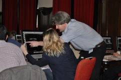
and Dr Alex Kirkham at University College London Hospital (UCLH) have successfully developed protocols to identify malignant tumours within the prostate gland using MRI. They use different modalities of MRI to produce the best anatomical data. The aim of the research is to evaluate if any MRI can be of use in predicting the future biological behaviour of the early prostate cancer.

MRI scans also allow the whole of the prostate to be examined. Previously, urologists believed that there were few tumours in the ‘front’ (anterior) of the prostate gland. This was because a 12 needle biopsy cannot reach this area. MRI and prostate mapping biopsies allow a thorough investigation of the whole prostate and precise treatment.

In addition to supporting research in this area – Pelican Cancer Foundation has also organised a series of Prostate Cancer Colloquiums where leading medical professionals can meet and discuss recent research into the various imaging techniques and their consequences for prostate cancer treatment.

Stay in touch

Sign up to hear from us
Donate Now

© 2023 Pelican Cancer Foundation - all rights reserved

  Registered Charity Number: 1141911 -  Privacy Policy